


Rat Spinal Cord Injury Model
 source:ELK Biotechnology
 date:2026-10-15
 views:60
Rat Spinal Cord Injury Model
1. Background
Spinal cord injury (SCI) may lead to loss of motor and sensory functions, and even death, bringing severe harm to patients. Multiple approaches are available to establish spinal cord injury models, including contusion, compression injury, traction dislocation, transection, ischemia, photochemical injury, excitotoxic injury and electrolytic injury models. Establishing stable rat spinal cord injury models provides standardized disease models for evaluating the efficacy of novel therapeutic interventions.
2. Experimental Materials and Methods
Experimental materials: SD rats (male, 180 220 g) Isoflurane (Manufacturer: RWD Life Science; Model: R510 22 10) Animal ventilator (Manufacturer: Shanghai ALCOTT Biotech; Model: ALC V8S) General small animal anesthesia machine (Manufacturer: RWD Life Science; Model: R580S)
Modeling procedure:
- Rats were acclimatized for 7 days before anesthesia.
- The bony landmark was identified at the boundary between the lowest rib and soft tissue on the dorsal side of rats. After surgical area disinfection, a 2.5 cm midline incision was made centered at the T10 segment to fully expose T9-T11 vertebrae. The spinous process and vertebral lamina of T10 were removed with rongeurs while keeping the dura mater intact. The T10 spinal cord was struck with an impactor. Successful model establishment was confirmed by immediate local hyperemia and edema of the injury site, together with bilateral hind-limb convulsions and tail swinging.
- Spinal cord tissues were collected 4 weeks post-surgery.
Evaluation indicators: Nissl staining, Hematoxylin Eosin (HE) staining.
3. Model Validation
Nissl staining
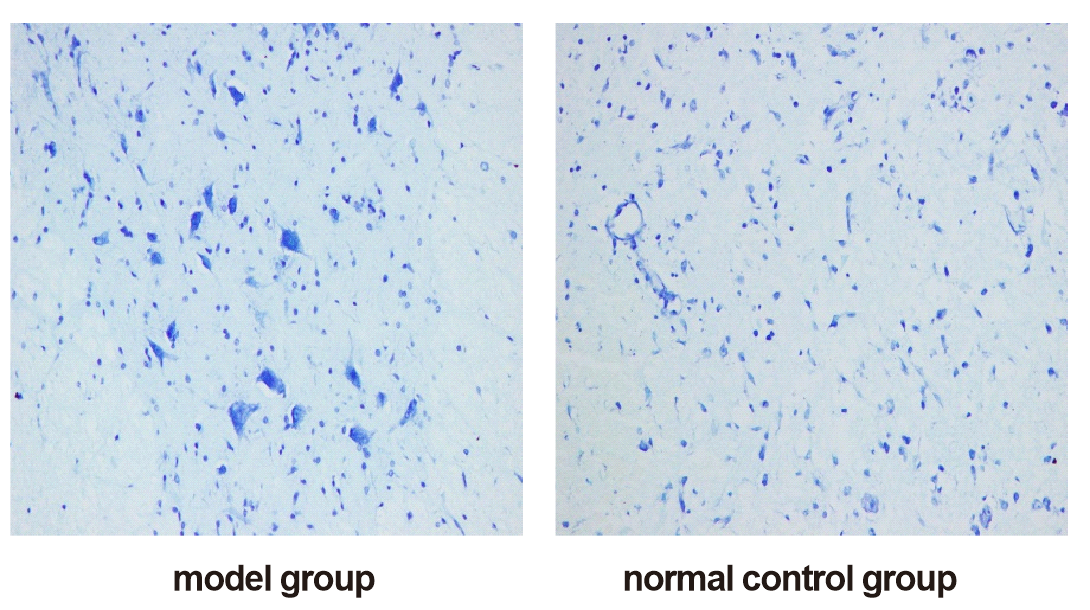
Normal group: Nissl bodies in spinal cord neurons were clearly visible; nuclei were large and round with distinct patch shaped Nissl bodies in cytoplasm. Model group: The number of Nissl bodies decreased with markedly weakened staining intensity; karyopyknosis was observed in partial fields of view, Nissl bodies became obscure, and inter neuron space increased.
HE staining
Normal group: The spinal cord tissue maintained basically intact structure; nerve cells were evenly distributed with normal morphology and clear Nissl bodies. Model group: Severe tissue hemorrhage and large hemorrhagic foci were observed with abundant blood cells in tissue; grey white matter structure was destructed and lacerated; partial nerve fibers dissolved and disappeared; tissue presented loose edema and vacuolar degeneration accompanied by reduced and lightly stained Nissl bodies.
Popular science Summary
Most clinical spinal cord injuries result from external contusions such as trauma impact and falls. Direct pathological mechanism research and new drug trials cannot be performed in human subjects. Therefore, researchers frequently establish rat spinal cord contusion models to mimic such human injuries. In this study, T10 segment of rat spinal cord was subjected to impact induced modeling to simulate traumatic spinal cord injury caused by external force. Nissl staining focuses on assessing the health status of neuronal cell bodies, whereas HE staining is applied to observe overall tissue hemorrhage, edema and structural damage. Comparison of stained sections reveals typical pathological changes after spinal cord injury, including hemorrhagic edema, laceration of nerve tissue structure and degenerative damage of neurons, which highly recapitulate pathological manifestations of human spinal cord injury. With this animal model, researchers can monitor disease progression and test the repairing efficacy of various drugs and interventions, laying an experimental foundation for clinical therapeutic research of spinal cord injury. Nevertheless, species differences exist between animal models and actual human injuries. Results obtained from animal experiments require further clinical validation and cannot be directly equivalent to therapeutic effects in human patients.

 RETURN
