


Three highly stable viral ELISA kits are now available!
 source:ELK Biotechnology
 date:2026-05-03
 views:678
Three highly stable viral ELISA kits are now available!
ELK is committed to premium quality, seamlessly aligning with your research pace.

Unveiling the Stealth Tactics of Influenza Virus: ELK's New ELISA Kits Target Key Viral Proteins
As seasons change, influenza viruses often make their quiet entrance. While most are familiar with the fever, aches, and fatigue they bring, behind these symptoms lies a precise molecular espionage campaign within the mucosal cells of our nose and throat. Today, ELK Biotechnology presents an overview of common human influenza virus classifications and detection methods, alongside introducing our latest products for influenza ELISA detection: the Human Influenza A Virus H3N2 ELISA Kit and the Human Influenza B Virus (NP) ELISA Kit.
I. The Four Major Families of Influenza Viruses
Influenza viruses belong to the Orthomyxoviridae family and are enveloped, single-stranded negative-sense RNA viruses. Their most distinctive feature is two key glycoproteins embedded in the viral surface: Hemagglutinin (HA) and Neuraminidase (NA). HA acts as the virus's "master key," responsible for recognizing and binding to sialic acid receptors on host cell surfaces. NA functions as the "cutting tool," severing the connection between the new virus particles and the host cell after replication to release progeny viruses.

The viral core consists of the RNA genome encapsulated by Nucleoprotein (NP). The antigenic specificity of NP forms the basis for typing influenza viruses. This sophisticated structure allows influenza viruses to efficiently invade cells while constantly mutating to evade immune surveillance.
Based on antigenic differences in NP and M1 proteins, influenza viruses are classified into four types:
| Type | Mutability | Host Range | Epidemic Features | Main Subtypes/Lineages |
| Type A (A) | Strongest; prone to antigenic drift and shift | Humans, birds, pigs, horses, etc. | Can cause seasonal epidemics and global pandemics | H1N1, H3N2, H5N1, H7N9, etc. (16 HA subtypes, 9 NA subtypes) |
| Type B (B) | Weaker; mutates relatively slowly | Primarily infects humans | Causes medium/small-scale epidemics or local outbreaks | Victoria lineage, Yamagata lineage (Note: Yamagata lineage has been scarcely detected since 2020) |
| Type C (C) | Stable; minimal mutation | Humans, pigs | Mostly causes sporadic cases with mild symptoms | No HA/NA subtype classification |
| Type D (D) | - | Cattle, pigs, and other livestock | Currently no evidence of pathogenicity to humans | - |
II. Mapping the Influenza Virus Invasion of the Human Body
The complete pathway of influenza virus invasion is a highly programmed molecular process:
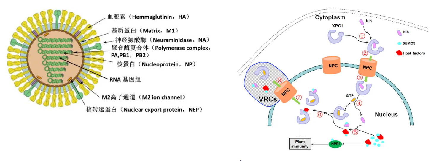
Step 1: Recognition and Attachment
The viral HA protein specifically binds to sialic acid receptors on the surface of respiratory ciliated epithelial cells. Human influenza viruses primarily recognize α-2,6-linked sialic acids (upper respiratory tract), whereas avian influenza viruses prefer α-2,3 linkages (lower respiratory tract).
Step 2: Endocytosis and Uncoating
Following binding, the cell internalizes the virus via receptor-mediated endocytosis, forming an endosome. Acidification of the endosome activates the viral M2 ion channel. A conformational change in HA mediates fusion between the viral envelope and the endosomal membrane, releasing the viral ribonucleoprotein complex (vRNP) into the cytoplasm.
Step 3: Replication and Transcription
The vRNPs enter the nucleus where the virus-encoded RNA-dependent RNA polymerase initiates replication and transcription. Viral mRNAs are transported to the cytoplasm for translation into viral proteins (HA, NA, NP, M1, etc.). These proteins then return to the nucleus to assemble with newly synthesized viral RNA into fresh vRNPs.
Step 4: Assembly and Release
Newly synthesized HA and NA are embedded into the host cell membrane. vRNPs bind to the matrix protein M1 and migrate beneath the cell membrane. Progeny viruses acquire their envelope by "budding" from the membrane. Finally, the NA enzyme cleaves sialic acids on the cell surface, releasing the new virus particles.
The entire replication cycle takes only 4-6 hours, demonstrating remarkable efficiency.
III. Common Detection Methods for Influenza Virus
1. Etiological Detection
Rapid Influenza Diagnostic Tests (RIDT): Results in 15-30 minutes. Simple to operate but cannot distinguish between subtypes.
Nucleic Acid Detection (PCR): One of the mainstream diagnostic methods. Offers high sensitivity and specificity, can differentiate virus types and subtypes. Typically requires 4-6 hours.
Viral Culture: A traditional method. Accurate but requires several days.
2. Serological Detection
Diagnosis by detecting influenza virus-specific antibodies (IgM/IgG) in blood. ELISA is a commonly used method.
IgM Antibodies: Rise 5-7 days post-infection, indicating recent infection.
IgG Antibodies: A fourfold or greater increase in titer during the convalescent phase compared to the acute phase has retrospective diagnostic significance.
IV. ELK Launches Two Precision ELISA Kits for Viral Detection

Building on a deep understanding of influenza virus biology, ELK Biotechnology introduces three ELISA kits targeting distinct and critical viral proteins, providing precise tools for your research:
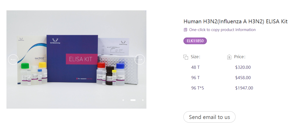
1. Human Influenza A Virus H3N2 ELISA Kit
Target: Influenza A Virus Nucleoprotein (NP)
Principle: NP is one of the most conserved proteins in the viral core, with far greater sequence stability than the surface hemagglutinin (HA). This kit specifically targets the NP protein of Influenza A H3N2 subtype (and broadly for Influenza A), effectively overcoming detection barriers posed by frequent surface antigen variation, offering exceptionally high detection specificity. Inter-assay precision CV% <10%.
Applications: Auxiliary diagnosis of Influenza A (H3N2), broad-spectrum epidemiological surveys across subtypes, dynamic monitoring of antibody levels post-infection.
Specification: 48T×5
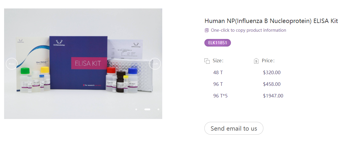
2. Human Influenza B Virus (NP) ELISA Kit
Target: Influenza B Virus Nucleoprotein (NP)
Principle: NP is one of the most conserved internal viral proteins, unaffected by surface antigen variation. This kit offers broad-spectrum recognition capability for both Victoria and Yamagata lineages of Influenza B virus, preventing false negatives due to surface protein variation. Inter-assay precision CV% <10%.
Applications: Diagnosis of Influenza B, epidemiological investigation, viral load quantification.
Specification: 48T×5
From virus classification to invasion mechanisms, from detection methods to precision tools—ELK Biotechnology not only provides detection kits but also aims to support your research journey with a comprehensive scientific perspective on influenza virology.
V. Product Recommendations
VI. Extended Product Offering
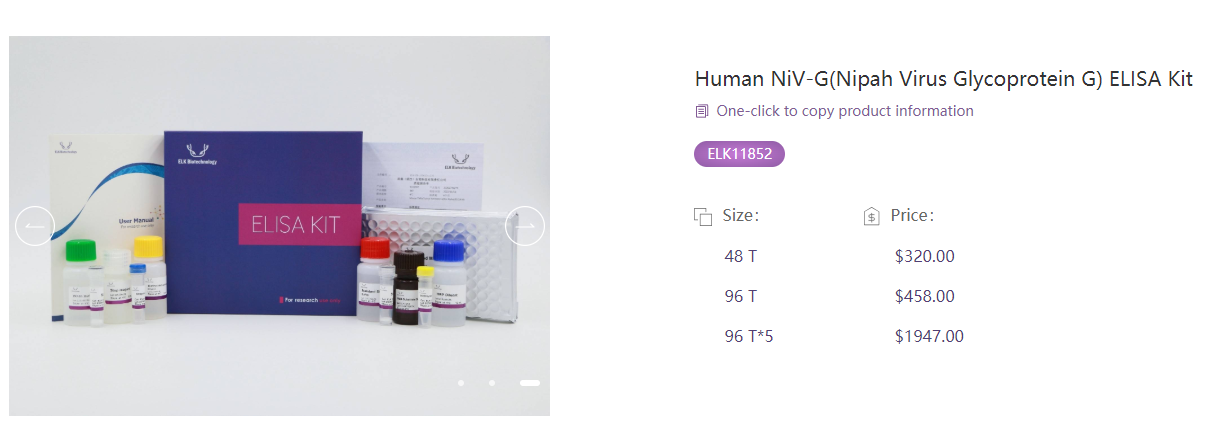
Human Nipah Virus Glycoprotein G (NiV-G) ELISA Kit
Target: Nipah Virus G Glycoprotein
Principle: Although Nipah virus belongs to the Paramyxoviridae family, its G protein functions similarly to influenza HA—mediating virus binding to host cell Ephrin-B2/B3 receptors. This kit provides an early serological diagnostic tool for this zoonotic disease with a fatality rate exceeding 40%. Inter-assay precision CV% <10%.
Applications: Early diagnosis of Nipah virus infection, antibody level monitoring, epidemiological studies.
Specification: 48T×5
Catalog Number: ELK11852

 RETURN