


A brief analysis of VEGF protein
 source:ELK Biotechnology
 date:2023-08-02
 views:389
The full name of VEGFA is vascular endothelial growth factor A.
Angiogenesis stimulators include a class of cytokines called “antigenic growth factors”, which play a role in stimulating cardiovascular formation. Some angiogenesis-stimulating factors have been found, such as platelet-derived growth factor (PDGF), fibroblast growth factor(bFGF), transforming growth factor (TGF),vascular endothelial growth factor A(VEGFA),etc. A key role in angiogenesis is VEGFA.
VEGFA protein is secreted by tumor cells, macrophages and fibroblasts, and is widely distributed in many tissues of the human body, such as glands, lungs, liver, kidney, myocardium, etc., but the expression level is extremely low, and its role is only to maintain normal blood vessel density and basic osmotic function to maintain nutrients. When tumor cells appear, their expression levels generally increase significantly. Studies have shown that VEGFA is associated with tumor invasiveness and tumor susceptibility.Gene Structure The VEGFA gene is located at position 6p12 of chromosome 6,with a full length of 16,272 bp, and there are seven splicing methods.
| mRNA | bp | exons | Protein | aa | exons |
| NM 001025366.1 | 3655 | 8 | NP 001020537.2 | 412 | 8 |
| NM 003376.4 | 3604 | 8 | NP 003367.4 | 395 | 8 |
| NM 001025367.1 | 3586 | 8 | NP 001020538.2 | 389 | 8 |
| NM 001025368.1 | 3532 | 7 | NP 001020539.2 | 371 | 7 |
| NM 001033756.1 | 3466 | 7 | NP 001028928.1 | 371 | 7 |
| NM 001025369.1 | 3497 | 7 | NP 001020540.2 | 354 | 7 |
| NM 001025370.1 | 3400 | 6 | NP 001020541.2 | 327 | 6 |
7 splicing methods of VEGFA
Gene molecular biology function
Vascular endothelial growth factor A(VEGFA) is the most important vascular endothelial growth factor(VEGF),In order to distinguish if from other vascular endothelial growth factor-related proteins, it is name after VEGFA. The VEGF family currently mainly includes VEGFA(ie VEGF),placental growth factor, VEGFB,VEGFC,VEGFD and VEGFD, among which VEGFA is an angiogenesis factor with the strongest and highest specificity in including tumor angiogenesis.
VEGFA is a high endothelial-specific mitogen that plays a major regulatory role in angiogenesis and formation. The reason why VEGFA has a high degree of endothelial cell specificity is that the protein has three high-affinity tyrosine kinase receptors(RTKs),VEGFR-1/Flt-1,VEGFR-2/KDR/Flk-1 and VEGF-3/Flt-4.KDR is the main regulatory molecule of angiogenesis, with obvious chemotactic and mitogenic effects, and is related to vascular island, angiogenesis and hematopoiesis;F1t-l mainly plays a role when endothelial cells align to form lumen. These two receptors are mainly expressed on endothelial cell, although very few hematopoietic cells and monocytes also have a small amount of expression, but only endothelial cells respond to VEGFA, so VEGFA is a growth factor that specifically acts on vascular endothelial cells.
The structure of VEGFA protein is a homodimer composed of two peptide chains through disulfide bonds. It is mainly produced by endothelial cells, mononuclear macrophages, and fibroblasts, it is commonly found in the central nervous systems, reproductive systems and tumor tissues. It’s main functions include:1,Selectively enhance mitosis of vascular endothelial cells, stimulate endothelial cell proliferation and promote angiogenesis.2,Increase the permeability of blood vessels, especially small blood vessels.so that plasma macromolecules are extravasated and deposited in the extravascular matrix, providing nutrients for the growth of tumor cells and the establishment of new capillary networks.
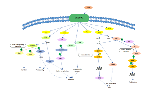
Pathways involved in VEGFA(VEGF)-mediated angiogenesis pathway map
VEGF plays an important role in angiogenesis, tumor growth, and ischemic disease. Hypoxia induced VEGFA expression in vitro. This may be because hypoxia-inducible factor-1(HIF1) acts on the hypoxia response element at the 5”end of VEGFA gene, which increase the transcription efficiency of VEGFA. The generated VEGFA protein binds to its receptor and induces angiogenesis through PKC,NOS,AKT,MAPK and other pathways.
Some VEGF related products provided by ELK Biotechnology
| Cat.No | Product name | price(48T/96T) |
| ELK1129 | Human VEGF ELISA Kit | 1680/2400 |
| ELK1293 | Mouse VEGF ELISA Kit | 1680/2400 |
| ELK2320 | Rat VEGF ELISA Kit | 1960/2800 |
| ELK1126 | Human VEGF R1/Flt-1 ELISA Kit | 1680/2400 |
| ELK1898 | Human VEGF R2/KDR ELISA Kit | 1960/2800 |
| ELK1287 | Mouse VEGF R2/FIK-1 ELISA Kit | 1680/2400 |
| ELK2256 | Human VEGF R3/Flt-4 ELISA Kit | 1960/2800 |
| ELK1194 | Human VEGF-C ELISA Kit | 1680/2400 |
| ELK1195 | Human VEGF-D ELISA Kit | 1680/2400 |
| ELK2321 | Human VEGF-B ELISA Kit | 1960/2800 |
The influence of genes on the occurrence of disease
VEGFA plays an important role in the pathogenesis of thyroid cancer. As the main driving factor of blood vessel growth, it can directly promote the growth of thyroid vascular endothelial cell or increase the permeability of blood vessels, and provide matrix for fibroblasts and endothelial cells, thereby promoting the formation of tumor blood vessels. In the 1970s,Folkman first proposed the theoretical hypothesis that tumor growth depends on cardiovascular generation in the ,<
Gene function research
In 1989,Ferrara et al. extracted VEGFA from bovine pituitary follicular stellate cells, which is a heparin-binding factor and is passed through two polypeptide chains with the same N-terminus and some differences in other regions. A highly conserved glycoprotein dimer linked by disulfide bonds with a molecular weight of 34-42KD.
In1992,Plate et al. believed that VEGFA could be expressed by tumor vascular endothelial cells, but not expressed in normal endothelial cells.
In 1994,Takahashi et al. Proved that VEGFA mainly exists in tumor cells, promots tumor angiogenesis, and has a synergistic effect with other angiogenesis-related factors(such as placental growth factor, basic fibroblast growth factor).
In 1995,Freeman et al. found that tumor-infiltrating lymphocytes could express VEGFA in the study of T lymphocytes.
In 1997,Masood et al. detected the synergistic high expression of VEGFA and its receptors Flt21 and KDR in AIDS Kaposi sarcoma cells, thus for the first time clearly proposed the autocrine mechanism of VEGFA in tumor cells.
In 1999,Ballamy et al. Studied hematopoietic malignancies(leukemia,lymphoma,and multiple myeloma) and found that there tumor cells not only expressed VEGFA, but also expressed VEGF receptors, suggesting that in addition to solid tumors, there is a In systemic malignancies, VEGFA also can play a role in the autocrine pathway.
In 2000,Dias et al. Demonstrated the autocrine mode of action of VEGFR-2-mediated growth and metastasis of human leukemia cells. Treatment of inoculated mice with the specific antibody IMC-C11 of human VEGFR-2(KDR) can block the interaction of KDR on VEGFA leukemia cells secreted by leukemia cells(it does not cross-react with murine VEGFA body Flt-1). This is ,blacking the autocrine mode of action, thereby hindering the growth of leukemia cells. It was proved for the first time that the VEGFA receptor expressed by leukemia cells is a functionally active signal transduction receptor, which transmits similar signals to endothelial cells.

 RETURN